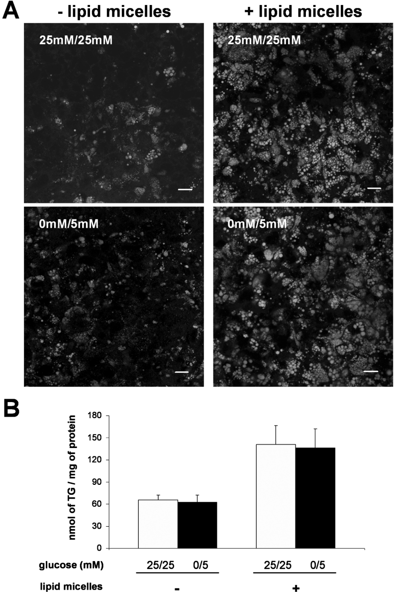
Figure 2. TG content of Caco-2 cells cultured in 25 mM/25 mM or 0 mM/5 mM glucose for 2 weeks post-confluence.
On the last day of culture, cells were supplied for 24 h with (right panels) or without (left panels) lipid micelles containing 2-MO. (A) Cells were stained for neutral lipids with BODIPY 493/503 and examined by confocal microscopy. The pictures shown are from the basal pole of the cells where cytosolic lipid droplets localize. Scale bar, 10 μm. (B) TG content in cell lysates. After extraction and separation by TLC, the TG spot was quantified using the PAP150 TG kit. Results shown, expressed as nmol of TG per mg of cell protein, are the means±S.E.M. for five independent experiments.

