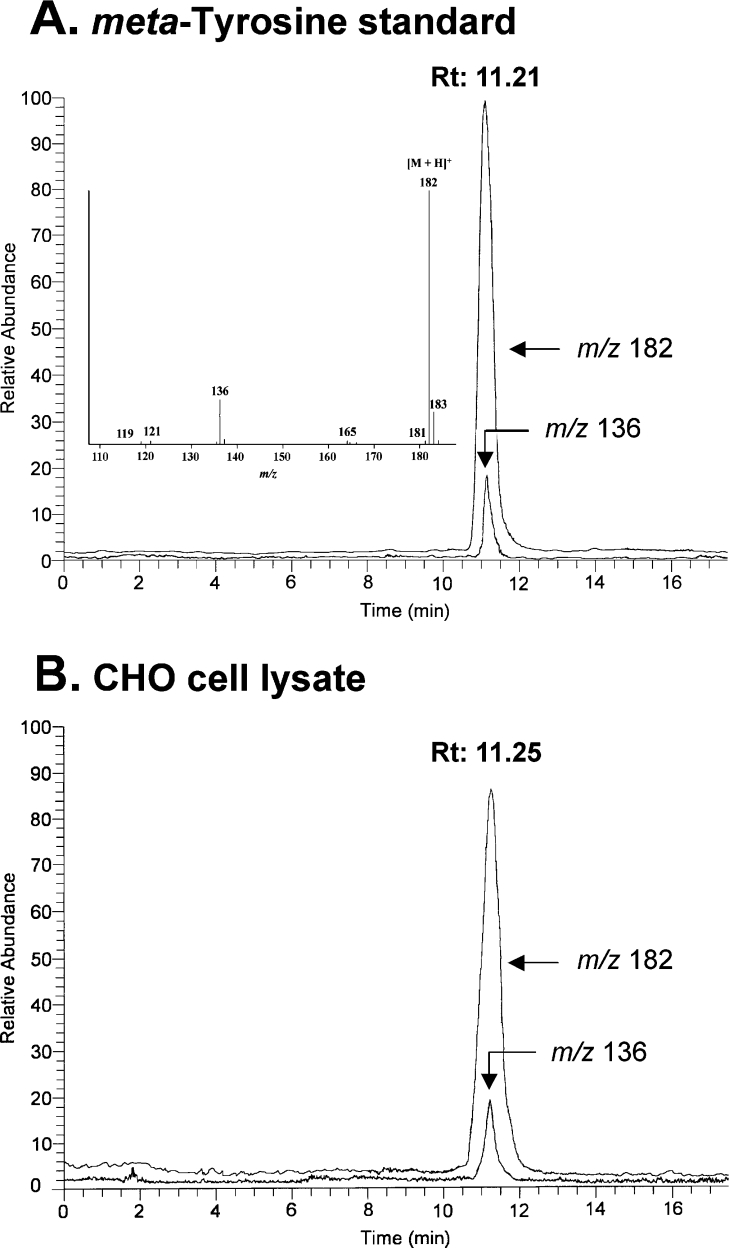
Figure 4. Detection of m-tyrosine in CHO cell proteins by positive-ion atmospheric pressure chemical ionization MS analysis.
(A) HPLC-MS analysis of authentic m-tyrosine subjected to HPLC on a reverse-phase column. Ions derived from the protonated molecular ion [M+H]+ and its major product ion [M−HCOOH+H]+ were monitored in the ion chromatogram. Note that the two ions co-elute and have the same relative abundance as in the full scan mass spectrum. The inset shows the mass spectrum of m-tyrosine (retention time 11.2 min). (B) HPLC-MS analysis of acid hydrolysates of CHO proteins subjected to HPLC on a reverse-phase column. Proteins were isolated from CHO cells incubated with medium A as described in Figure 2. Note that the relative abundance of the ions (m/z 182 and m/z 136) and retention times (Rt; 11.21 and 11.25 min) of authentic m-tyrosine and the material in hydrolysates of CHO proteins are virtually identical.

