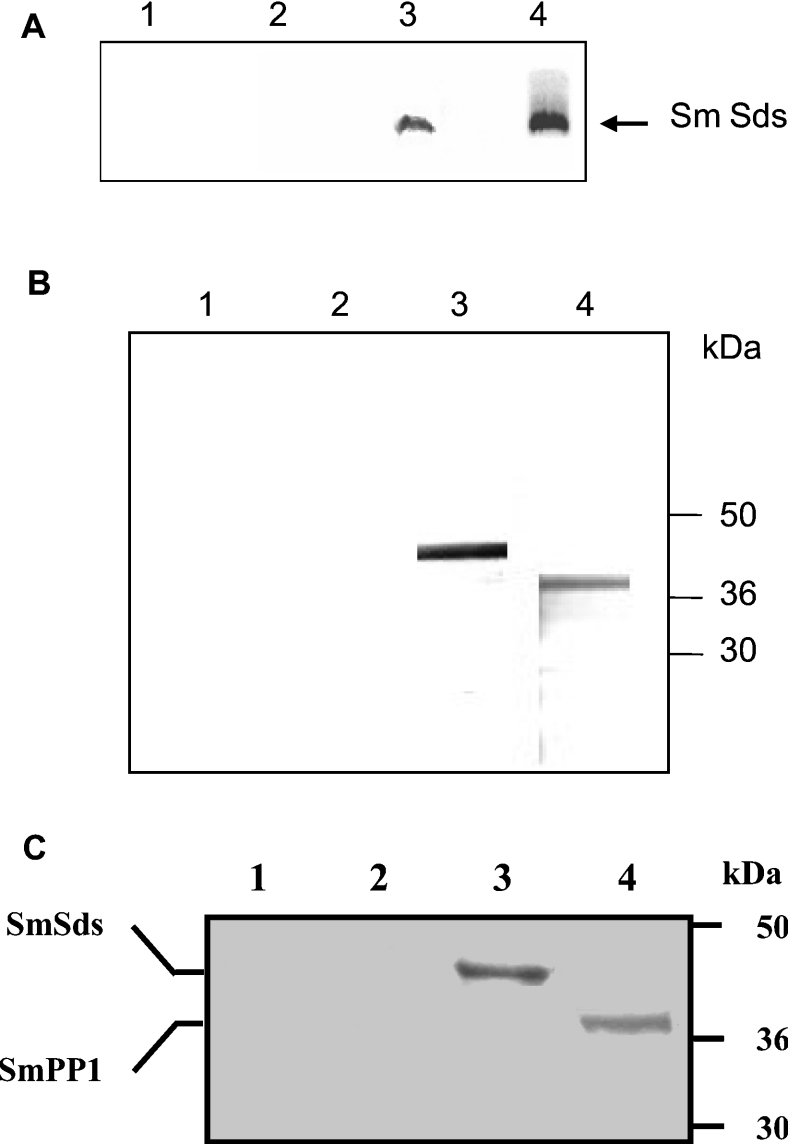
Figure 5. Interaction of SmSds with SmPP1 in vitro and in Schistosoma mansoni.
(A) GST pull-down assays were performed with 35S-labelled SmSds incubated with beads alone (lane 1), GST bound to beads (lane 2) and GST–PP1-bound beads (lane 3). Lane 4 represents 20% of the input. (B) Immunoblot analysis of proteins eluted from microcystin–agarose. Eluates were separated by SDS/PAGE (4–12% gels) and transferred on to nitrocellulose. Lanes 1 and 2 represent the negative controls (pre-immune rat and mouse sera respectively). Lanes 3 and 4 represent the detection of SmSds and the detection of SmPP1 respectively. (C) Co-immunoprecipitation of the SmSds–SmPP1 complex with anti-SmSds antibodies from Schistosoma mansoni extracts. Immunoprecipitates from control pre-bleed sera or from SmSds antisera were eluted, separated by SDS/PAGE (4–12% gels) and transferred on to nitrocellulose. Immunoblot analysis was performed with anti-SmSds (lanes 1 and 3) and anti-SmPP1 (lanes 2 and 4) antibodies. Molecular-mass sizes are given in kDa.

