
Figure 7.
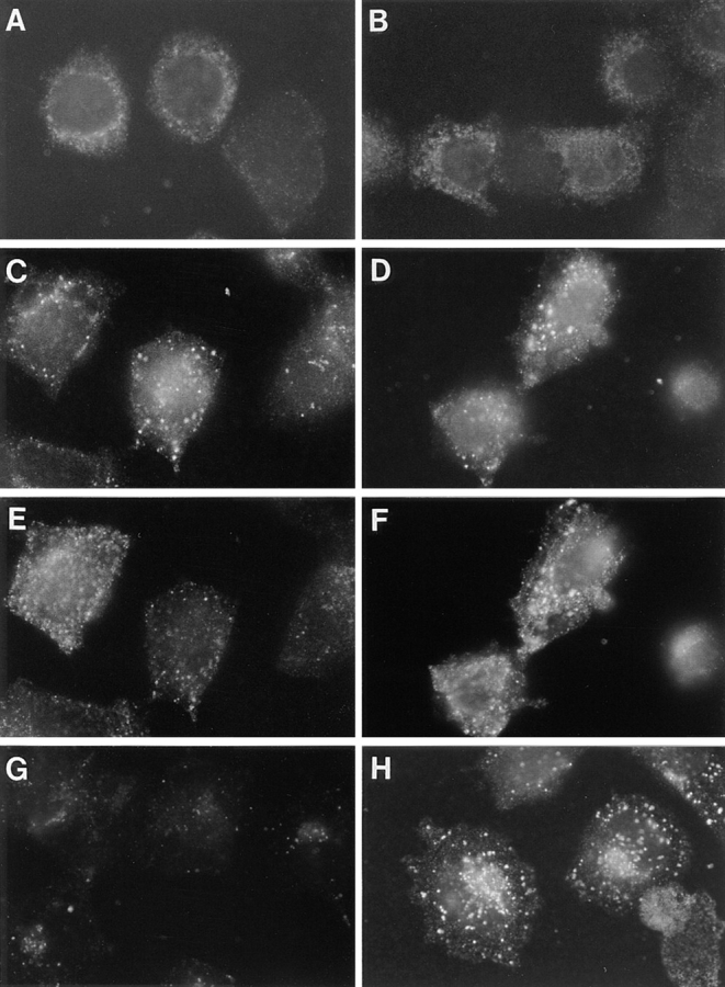
Time course of fur/f internalization. Parallel plates of M2 (A, C, E, and G) and A7 (B, D, F, and H) cells grown on glass coverslips were infected with VV:hfur/f (moi = 10). At 4 h after infection, mAb M1 (6 μg/ml final concentration) was added to the culture media for either 5 min (A and B), 15 min (C–F) or 60 min (G and H). Cells in C–F were also incubated with 40 ng/ml r-Tf and 100 nM tautomycin (to accumulate internalized furin in the early endosomes). C and D show mAb M1 and E and F show r-Tf in double-labeled cells. Samples were processed for immunofluorescence microscopy as described in Fig. 5. Internalized mAb M1 was visualized with anti–mouse IgG2b-TXR antibody.