
Figure 9.
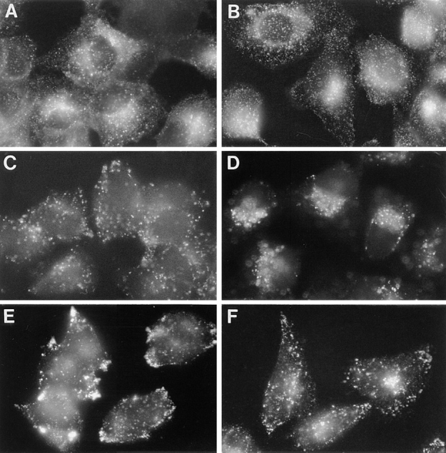
Importance of ABP-280 to the localization of endocytic compartments. Parallel plates of M2 (A, C, and E) and A7 (B, D, and F) cells were grown on coverslips. Early endocytic compartments were visualized by treating the cultures with rhodamine-transferrin (r-Tf, 40 ng/ml) for 30 min at 37°C before fixation (A and B). Late endosomes were visualized by addition of Texas Red-dextran beads (10,000 mol wt, 10 μg/ml) for 30 min at 37°C before fixation (C and D). To visualize lysosomes, cells were fixed, permeabilized with detergent, and incubated with a LAMP-1 antibody followed by incubation with anti–mouse IgG1-FITC (E and F).