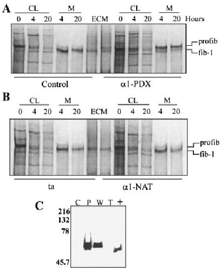
Fig. 4.

Fibroblast cell strains infected with adenovirus constructs expressing α1-PDX demonstrated delayed processing of profibrillin-1 to fibrillin-1. Fibroblasts were infected with transactivating virus (ta) either by itself, or co-infected with α1-PDX or α1-NAT. The cultures were radiolabeled with [35S]cysteine for 30 min, followed by both 4 and 20 h chases with non-radioactive DMEM. CL and media (M) fractions were harvested and separated by SDS–PAGE. A: Uninfected control cultures (control) and cultures co-infected with ta and α1-PDX (α1-PDX). B: Cultures infected with ta alone (ta) and cultures co-infected with ta and α1-NAT (α1-NAT). The positions of profibrillin-1 (profib-1) and fibrillin-1 (fib-1) are indicated. C: Immunoblot analysis of the media from infected cultures. Media was separated by SDS–PAGE through 10% gels and transferred to PVDF. Immunoblot analysis with mAb M2 detected positive signals in α1-PDX and α1-NAT infected CL and media. C, control; P, infected with α1-PDX and ta; W, infected with α1-NAT and ta; T, infected with ta.
