Fig. 5.

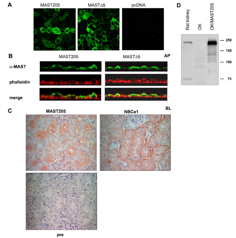
Subcellular localization of MAST205. A: monolayers of transfected OK cells were fixed and stained with anti-MAST205 antiserum followed by Alexa Fluor 488-conjugated secondary antibody (green). Focal plane views of MAST205-, MΔ5-, or pcDNA3.1HisB-transfected cells are shown. B: vertical sectional views of MAST205- or MΔ5-transfected cells are shown. Actin was labeled with phalloidin (red) to show the location of the apical (AP) and basolateral (BL) membrane. The presence of MAST205 and MΔ5 at the apical membrane is evident as yellow fluorescent signals, indicating the colocalization of MAST205 and actin. C: immunohistochemical staining of rat renal proximal tubules with the anti-MAST205, anti-NBCe1, or preimmune (pre) serum. MAST205 is expressed at the brush-border membrane. As a control, the basolateral staining of NBCe1 is shown. No staining with the preimmune serum was observed. D: Western immunoblot using the anti-MAST205 antibody on lysates prepared from rat kidney, OK, and OK cells transfected with mouse MAST205.
