Figure 10.
Immunolocalization of LPA1 and Light-Induced Accumulation of LPA1 and Plastid Proteins.
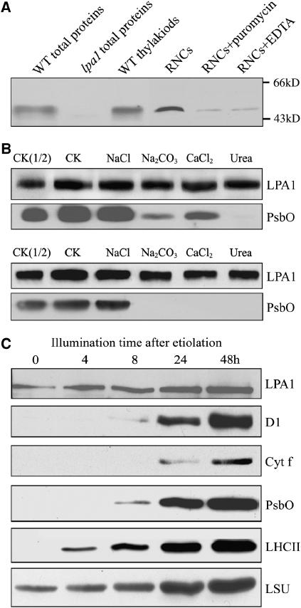
(A) Immunoblot analysis of LPA1. Samples from wild-type and lpa1 plants consisting of total leaf proteins, the thylakoids (equivalent to 5 μg chlorophyll), RNCs and RNCs treated with 1 mM puromycin or 10 mM EDTA (equivalent to 50 μg chlorophyll) were separated by SDS-PAGE and immunodetected with the antibody raised against LPA1.
(B) Salt washing of the membranes. The membrane preparations were incubated with 1 M NaCl, 200 mM Na2CO3, 1 M CaCl2, and 6 M urea for 30 min at 0°C (top panel) or sonicated in the presence of these salts (bottom panel) and incubated for another 30 min at 0°C. PsbO, the 33-kD luminal protein of PSII, was used as a marker. CK, the membrane preparations without treatments were used as a control.
(C) Immunoblot analysis of protein accumulation during light-induced greening of etiolated Arabidopsis seedlings. After growth in the dark for 5 d, the etiolated seedlings were illuminated for 4, 8, 24, and 48 h. The samples were harvested and processed for immunoblot analysis. Protein loadings were based on equal amounts of seedling fresh weight. The antibodies used for the analysis are indicated at the right.

