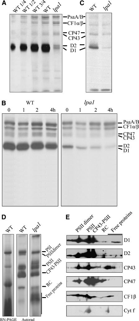
Figure 6.
In Vivo Protein Synthesis of Plastid-Encoded Membrane Proteins and Immunoblot Analysis after Second Dimension BN-PAGE.
(A) Incorporation of [35S]Met into thylakoid membrane proteins in young seedlings. After a 20-min pulse in Arabidopsis young seedlings in the presence of cycloheximide, the thylakoid membranes were isolated, and the proteins were separated by SDS-urea-PAGE and visualized autoradiographically.
(B) Pulse and chase of thylakoid membrane proteins. The 20-min pulse in Arabidopsis young seedlings was followed by a chase of cold Met for 1, 2, and 4 h. The thylakoid membranes were then isolated, separated by SDS-urea-PAGE, and visualized autoradiographically.
(C) Incorporation of [35S]Met into thylakoid membrane proteins in mature leaves. After a 30-min pulse in Arabidopsis mature leaves in the presence of cycloheximide, the thylakoid membranes were isolated, and the proteins were separated by SDS-urea-PAGE and visualized by autoradiogram.
(D) Autoradiogram of thylakoid membrane protein complexes of young seedlings solubilized and separated by BN-PAGE after pulse labeling for 20 min. A lane of BN gel separated thylakoid membrane proteins is shown at the left to indicate the locations of the identified complexes. RC, reaction center.
(E) Representative immunoblots from a second dimension SDS-PAGE separation of the thylakoid membrane. Horizontal strips of immunoblots with anti-D1, anti-D2, anti-CP47, anti-CP43, anti-CF1β, and anti-cytochrome f antibodies are shown.

