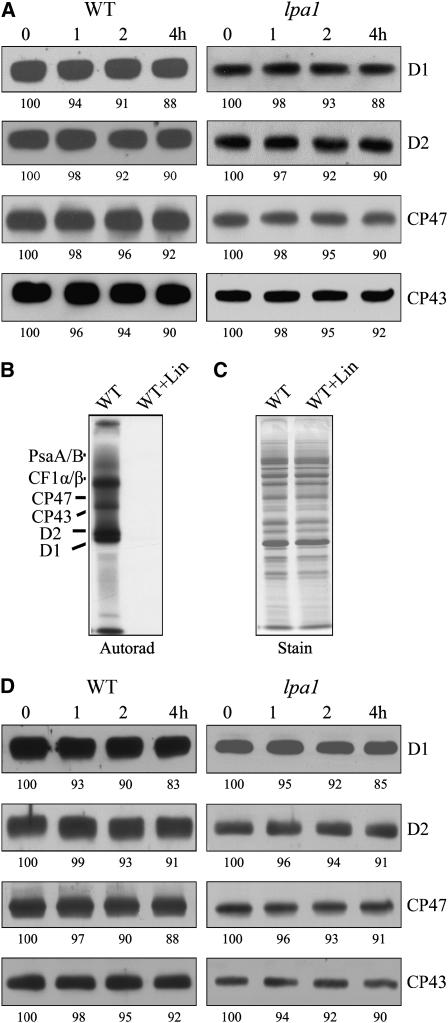
Figure 7.
Stability of PSII Proteins.
(A) Immunoblot analysis of thylakoid membrane proteins. The Arabidopsis leaves were incubated with lincomycin for 30 min and illuminated for various times. After this treatment, the thylakoid membranes were isolated, and the contents of PSII proteins were determined through immunoblot analysis. X-ray films were scanned and analyzed using an AlphaImager 2200 documentation and analysis system. The percentages of protein levels shown below the lanes were estimated by comparison with levels found in corresponding samples taken at time 0.
(B) and (C) Effects of lincomycin (Lin) on protein synthesis. After incubation in the presence of 20 μg/mL cycloheximide and 100 μg/mL lincomycin for 30 min, the Arabidopsis leaves were labeled for 20 min. After labeling, the thylakoid membrane proteins were isolated, separated by SDS-urea-PAGE, and visualized autoradiographically (B). The Coomassie blue–stained gel (C) is presented to show that equal amounts of proteins were loaded.
(D) Immunoblot analysis of thylakoid membrane proteins. The Arabidopsis leaves were incubated with cycloheximide for 30 min and illuminated for various times. After the treatments, the thylakoid membranes were isolated, and the contents of PSII proteins were determined through immunoblot analysis. The percentages of protein levels shown below the lanes were estimated by comparison with levels found in corresponding samples taken at time 0.

