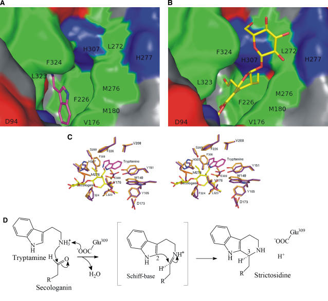
Figure 4.
Surface Representation of STR1-Ligand Complexes.
Hydrophobic residues (Tyr, Trp, Phe, Leu, Met/Mse, Cys, Ile, and Val), positively charged residues (Arg, Lys, and His), negatively charged residues (Asp and Glu), and hydrophilic residues (Ala, Gly, Ser, Thr, Pro, Gln, and Asn) are shown in green, blue, red, and gray, respectively. The surrounding residues are labeled with single-letter codes.
(A) Close-up view of the substrate binding pocket with tryptamine in stick representation.
(B) Close-up view of the substrate binding pocket with secologanin in stick representation.
(C) Stereoview of tryptamine and secologanin together and superposition of the surrounding residues. The color code is as described in Figure 3.
(D) Schematic presentation of the reaction pathway for the Pictet-Spengler–type reaction with Glu-309 involved in the amine deprotonation.

