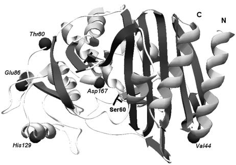
FIG. 1.
Predicted 3D model of the AmpC β-lactamase from M. morganii strain PP29, constructed using Swiss-Model and the Deep-View/Swiss-Pdb Viewer v3.7. The following amino acid shifts, compared to AmpC GUI-1 (AF055067), are shown as dark spheres: Ile44Val, Ala80Thr, Ala86Glu, Asn129His, and Glu167Asp. The active-site serine residue (Ser60) is also shown. N, amino terminal; C, carboxy terminal.

