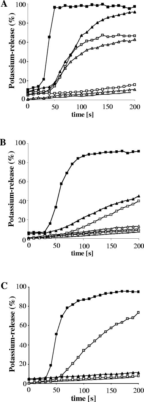
FIG. 3.
Potassium release from Micrococcus flavus DSM 1790 (A), Staphylococcus simulans 22 (B), and Lactococcus lactis HP (C) induced by gallidermin (triangles) and nisin (squares). Peptides were added at 30 seconds, and the potassium release was monitored with a potassium electrode. Potassium leakage is expressed relative to the total amount of potassium released after addition of 1 μM nisin (100% value). Peptides were applied at 500 nM (black symbols), 50 nM (gray symbols), or 5 nM (white symbols). Lines without symbols are baselines.

