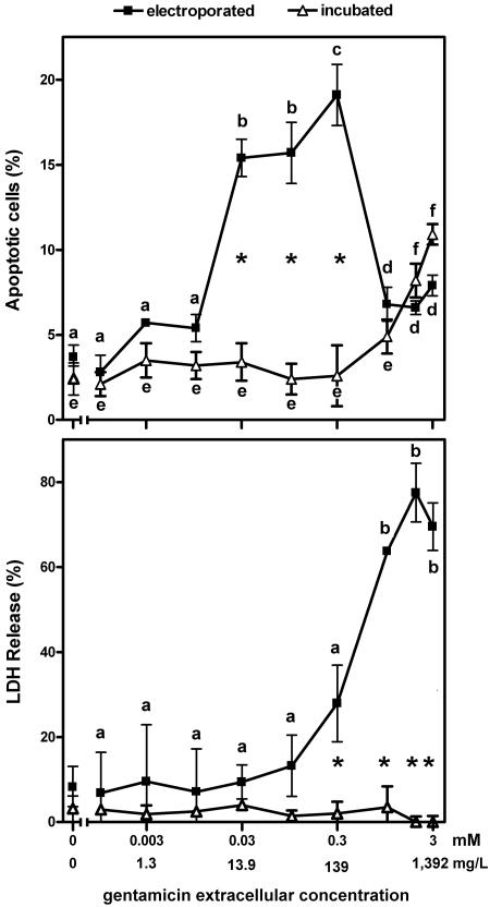
FIG. 3.
Influence of gentamicin concentration on apoptosis (upper panel) and necrosis (lower panel) in LLC-PK1 cells. Apoptosis was assessed by enumeration of typical apoptotic nuclei (DAPI staining; see Fig. 1). Necrosis was assessed by measuring the release of the cytosolic enzyme lactate dehydrogenase (LDH) in the culture medium (shown as the percentage of the total amount in cells plus medium). Electroporated, cells were subjected to electroporation in the presence of gentamicin at the concentrations showed in the abcissa, returned to gentamicin-free medium, and examined 24 h later; incubated, cells were cultivated for 24 h in the presence of gentamicin at the concentrations shown on the abscissa. The 0 value on the abscissa corresponds to cells electroporated (closed symbols) or incubated (open symbols) in the absence of gentamicin. Values are means ± standard deviations (n = 3). Statistical analysis was performed by the use of analysis of variance, and the values of the datum points with different letters in the electroporated cell or the incubated cell panels are significantly different from those of all other points in the same group (P < 0.05; for lactate dehydrogenase release [lower panel], there is no significant difference among any of the datum points for incubated cells); the asterisks show the significant differences (P < 0.05) between groups (electroporated versus incubated cells).

