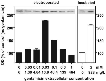
FIG. 5.
Detection of the proapoptotic protein Bax by Western blot analysis of lysates of LLC-PK1 cells. The films were analyzed by densitometry (with measurements done in triplicate; the values shown are means ± standard deviations; when not visible, the standard deviation bars are smaller than the minimal resolution of the graph). Electroporated (left panel), cells were subjected to electroporation in the presence of gentamicin at the concentrations shown on the abscissa and returned to drug-free medium for 8 h at 37°C before they were collected and processed; incubated (right panel), cells were maintained at 37°C for 8 h in the presence of gentamicin at the concentrations indicated on the abscissa. The 0 value on the abscissa corresponds to cells electroporated (closed bars) or incubated (open bars) in the absence of gentamicin. OD, optical density.

