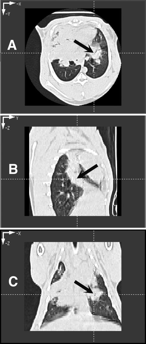
FIG. 2.
CT images of rabbit lung in three planes. (A and B) Transverse plane depicting pulmonary infiltrates in both lower lobes (A) and section of lungs depicting pulmonary infiltrates due to invasive pulmonary aspergillosis (B). Note that the pulmonary infiltrate when viewed in the sagittal plane depicts a column of infiltrate, which on transverse section appeared to be spherical. (C) Coronal image plane of rabbit lungs with experimental invasive pulmonary aspergillosis depicting irregularly contoured pulmonary infiltrates.

