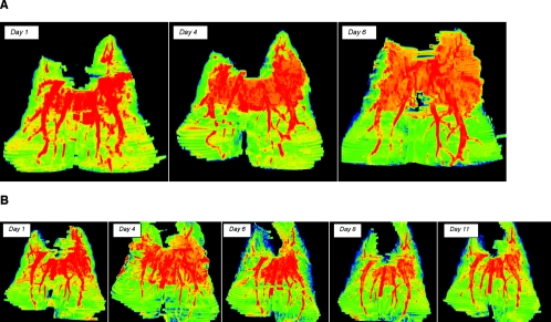
FIG. 3.
Pseudocolor images of multidimensional reconstruction of pulmonary infiltrates of experimental aspergillosis. (A) Pseudocolor images of pulmonary infiltrates due to experimental pulmonary aspergillosis of untreated controls demonstrate progressive lesions expanding in three dimensions in predominantly the upper lobes. Day 1 images demonstrate highly dense infiltrate in association with early pulmonary hemorrhage; as the lesion expands, there is an increasing level of infection with progressive blood vessel involvement due to angioinvasion. By day 6, the lesions have expanded and have changed in density, reflecting the development of pulmonary infarcts, as well as disruptive blood vessels in association with angioinvasion. (B) Multidimensional pseudocolor reconstruction of experimental pulmonary aspergillosis in a rabbit treated with deoxycholate amphotericin B. The reconstructed CT image on day 1 depicts early pulmonary hemorrhage and expanding infiltrates. However, by day 4 infiltrates have stabilized and have tended to diminish. There is early evidence of blood vessel invasion. By day 6, there are substantial resolution of infiltrates and an apparent increase in vessels, which continue into day 8 and finally day 11. By day 11, pulmonary infiltrates have virtually resolved and the blood vessels have returned to a normal anatomic structure.

