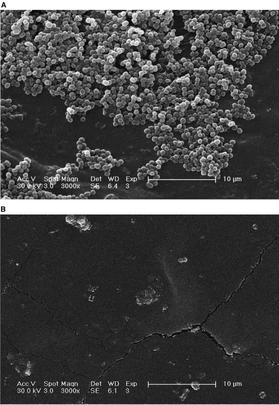
FIG. 6.
(A) Scanning electron micrograph of the surface of a section of a Lubri-sil hydrogel-coated all-silicone Foley catheter after biofilm formation by S. epidermidis 414 for 24 h (×3,000 magnification). Attached cells are clearly visible in large quantities on the surface. (B) Surface of a section of a Lubri-sil hydrogel-coated all-silicone Foley catheter pretreated with phage 456 after biofilm formation by S. epidermidis 414 for 24 h (×3,000 magnification) with divalent cation-supplemented MHB. No biofilm matrix or clusters of attached cells are visible (images represent typical fields of view).

