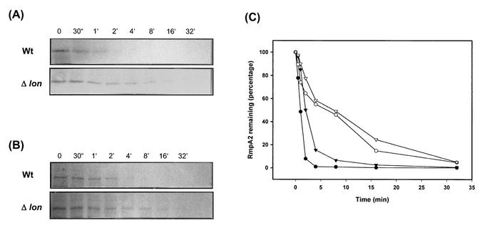
FIG. 6.
Stability of RmpA2 protein in K. pneumoniae. The bacterial cells were pulse-labeled with [35S]methionine and chased at the indicated time points. Tag-fused RmpA2 protein was immunoprecipitated with anti-His MAb or with anti-HA MAb and then subjected to SDS-PAGE. (A) Turnover of His-RmpA2 protein. (B) Turnover of HA-RmpA2 protein in the wild-type strain K. pneumoniae CG43S3 (Wt) or in the lon mutant strain L2117 (Δlon). (C) Quantification of the autoradiogram shown in panel A of wild-type (solid circles) or lon mutant (open circles) cells and of that in panel B of wild-type (solid triangles) or lon mutant (open triangles) cells. The quantity of labeled protein at time zero was set at 100%.

