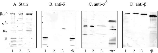
FIG. 1.
RNAP purification and subunit analysis. A total of 1 to 2 μg of purified RNAP from GBS strains A909 (1) and AJ200 (2) and B. subtilis strain MH5636 (3) were separated on a 12% SDS-polyacrylamide gel and stained with Coomassie blue (A). Subunit identity is shown on the left. Western blot analysis was performed on the RNAP preparations by using anti-δ (B), anti-σA (C), or anti-β (D) antisera. Recombinant δ (rδ), σA (rσA), and β (rβ) served as positive controls. Immunoreactive bands were detected by using an infrared imager after incubation with a rabbit anti-IgG infrared-labeled secondary antibody.

