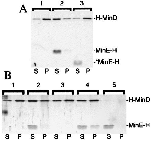
FIG. 6.
Stabilization of a MinE-MinD-membrane complex in the presence of ATPγS. SYPRO Ruby-stained gels showing the effect of MinE-H on, and the interaction of MinE-H with, prefractionated H-MinD-membrane complexes are shown. (A) Vesicles were decorated with H-MinD.ATP and treated with buffer (lane 1), MinE-H (lane 2), or *MinE-H (lane 3) as described in the legend to Table 4. (B) Vesicles were incubated with H-MinD (lanes 1 to 4) or without (lanes 5) and in the presence of either ATP (lanes 1 and 2) or ATPγS (lanes 3 to 5). Vesicles were harvested by centrifugation and resuspended in RB with the same nucleotide, and either 1 μM MinE-H (lanes 2, 4, and 5) or the same volume of buffer (lanes 1 and 3) was then added. After incubation, mixtures were refractionated, and equivalent aliquots of the supernatant (S) and pellet (P) fractions were subjected to SDS-PAGE. For additional details, see the text and the legend to Fig. 7.

