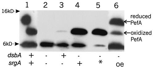
FIG. 4.
Western immunoblot analysis of SrgA dependence of oxidation status of PefA. Blot was developed with anti-PefA antibodies. Half of the PefA protein in lane 1 shifted upwards to run at the reduced location as a result of dithiothreitol diffusion from the markers in the adjacent lane (not shown). Lane 1, NLM2153/p30; lane 2, NLM2198/p30Tn2.7; lane 3, NLM2153/p30Tn2.7; lane 4, NLM2198/p30; lane 5, NLM2198/p30Tn2.7/pCB6; lane 6, NLM2198/p30Tn2.7/pCB6 with arabinose induction. The presence or absence of dsbA and srgA is indicated at the bottom of the figure (* indicates the presence of srgA in trans; oe indicates overexpression of srgA). The anti-PefA antibody cross-reacts with material running at the dye front of the gel (6-kDa marker). The relative positions of the 16- and 6-kDa molecular size markers are indicated.

