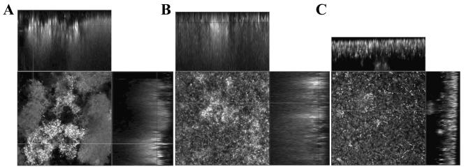
FIG. 3.
Confocal laser scanning microscopic images of 6-day-old biofilms of Streptococcus pneumoniae serotypes. Representative CLSM images of S. pneumoniae group I biofilm architecture (A) of strain BS71 (serotype 3), group II biofilm architecture (B) of strain BS73 (serotype 6), and group III biofilm architecture (C) of strain BS75 (serotype 19) are shown. Biofilms were grown in flow cells under once-through flow conditions for 6 days, after which time the biofilms were stained with the Live/Dead BacLight stain. Biofilms were viewed at 400× magnification. The CLSM images show the xy and xz planes. Flow cell experiments were performed in triplicate as described in Materials and Methods.

