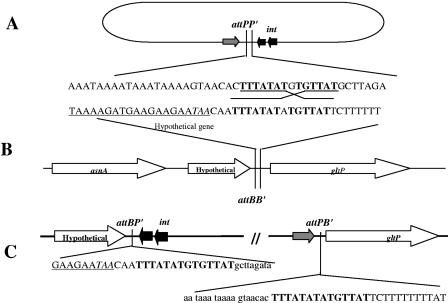
FIG. 5.
Organization of bacterial and phage attachment sites. (A) Schematic representation of circularized phage genome with its attPP′ site and nearby genes. (B) C. difficile genome showing attBB′ site and surrounding genes. (C) Partial sequences of junctions showing the phage sequence in lowercase letters, the bacterial sequence in uppercase letters, and the homologous att site in boldface letters. The underlined sequence is the 3′ end of the hypothetical gene, and the stop codon is in italics.

