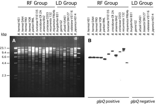
FIG. 4.
Distribution of glpQ among Borrelia species demonstrated by Southern blot analysis. (A) Agarose gel with genomic DNA digested with EcoRI and stained with ethidium bromide. (B) Hybridization pattern with the glpQ probe, which was identical to the hybridization pattern with the glpT probe (data not shown). Molecular size standards in kilobase pairs are on the left. The relapsing-fever (RF) spirochetes were positive with both probes, while the Lyme disease (LD) group of spirochetes were negative. All samples hybridized with the gpsA probe (data not shown).

