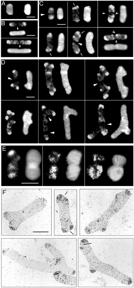
FIG. 2.
Segregation of murein in d-cysteine-labeled sacculi from E. coli CS801-4. Cells labeled with d-cysteine were transferred to d-cysteine-free LB medium with and without 1 μg of aztreonam per ml. Samples were removed when the OD550 increased by three- and fivefold, and murein was purified and processed for immunodetection of d-cysteine-containing and total murein as described in the text. As a control, a culture of the parental strain CS109 was d-cysteine labeled and chased in parallel. Samples were removed at the initiation of the chase period and when the OD increased by threefold. Sacculi were observed either by confocal (A to E) or by electron (F) microscopy. Fluorescence pictures were captured in two channels; one would image the distribution of d-cysteine-containing murein (left or upper image in each frame), and the second would image total murein (right or bottom image in each frame). Mosaics depicting selected sacculi were constructed with Adobe Photoshop software. (A) CS801-4 sacculi at the initiation of the chase period. (B) Sacculi from CS109 chased in the presence of aztreonam for a threefold increase in OD. (C) Sacculi from CS801-4 chased in the presence of aztreonam for a threefold increase in OD. (D) Sacculi from CS801-4 chased in the presence of aztreonam for a fivefold increase in OD. (E) Sacculi from CS801-4 chased in the absence of aztreonam for a threefold increase in OD. (F) Sacculi from CS801-4 chased in the presence of aztreonam for a fivefold increase in OD. Silver grains reveal areas of d-cysteine-containing murein. The electron microscopic negatives were digitized and further processed for the mosaic picture with Adobe Photoshop software. Bars, 2.5 μm. Triangular arrowheads indicate potential division sites made up of “all new” murein, V-shaped arrowheads indicate regions of conserved murein outside the poles, and small arrows indicate apparently split poles.

