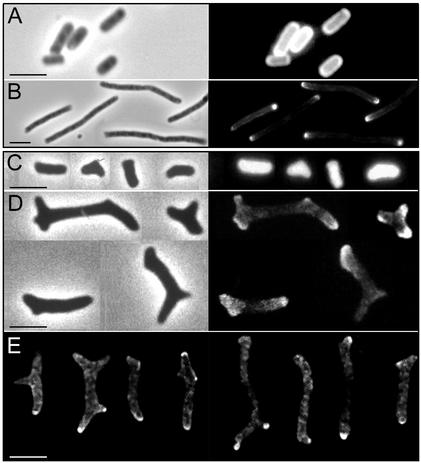
FIG. 3.
Segregation of Texas Red succinimidyl ester-labeled surface components. Cells of CS801-4 and its parental strain CS109 were labeled with Texas Red X-succinimidyl ester, transferred to LB medium plus 1 μg of aztreonam per ml, and incubated at 37°C. Samples were removed immediately after dilution and when the OD had increased by fivefold. Cells were fixed and further processed for phase-contrast/epifluorescence or confocal microscopy as described in the text. (A) Unchased cells of CS109; (B) chased cells of CS109; (C) unchased cells of CS801-4; (D) chased cells of CS801-4. Right panels show the fluorescence image for Texas Red in the cells visualized by phase contrast in the left panels. (E) Selected cells of CS801-4 as observed by confocal microscopy. The optical planes showed are those apparently corresponding to the central sections of the cells. It is relevant that CS801-4 cells do not extend flat on the glass slides because they are bent in more than one plane. Nominal thickness of the optical sections was 0.53 μm. Bars, 5 μm. The figure was assembled from digitized photographic slides (phase contrast and epifluorescence) or from digital images (confocal) with Adobe Photoshop software.

