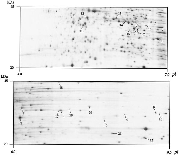
FIG. 3.
Analyses of B. pertussis BPDR proteins by 2-D electrophoresis. The portions of pH 4 to 7 (top) and pH 6 to 9 (bottom) silver-stained gels corresponding to the 20- to 45-kDa range are shown. Only the putative periplasmic proteins involved in solute binding as identified by mass fingerprinting have been numbered (1 to 6, Bug9, -2, -73, -20, -71, and -4, respectively; 7 to 10, predicted TRAP transporter-associated ESRs; 11 to 22, predicted ABC transporter-associated ESRs). See http://www.ibl.fr/articles/jbact_antoine.htm (Table A3) for the sequences of these 22 proteins.

