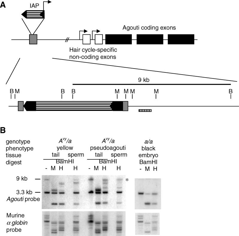
Figure 1. Methylation of the Avy Allele in Mature Sperm.
(A) Expression of the Avy allele is controlled by an IAP, inserted into pseudoexon 1a (grey box). A cryptic promoter within the 3′ LTR of the IAP (black arrows) directs transcription of the agouti coding exons. The BamHI (B) and MspI (M) sites are shown in the region of the unique 400-bp probe B. Tail and mature sperm from a yellow and a pseudoagouti male were collected. DNA was prepared and samples digested with BamHI followed by MspI or its isoschizomer HpaII, transferred and hybridised with the agouti probe [3]. The Avy allele produces a 9-kb BamHI band, while the a allele produces a 3.3-kb band. Membranes were stripped and rehybridised with a murine α-globin probe to check for equal digestion within the tissue samples (shown in [B]). These results represent experiments performed on sperm and tail DNA from seven yellow and five pseudoagouti males, a further one of each are shown in Figure S1. Mature sperm were isolated from both epididymes of the male (each sample contained in the order of 106 to 107 spermatocytes). Sperm samples were checked by light microscopy and found to be greater than 95% spermatocytes. The methylation state of the tissues is indicated by the ratio of the 9-kb BamHI band to the 7-kb band remaining after HpaII digestion. The 9-kb band is marked by an asterisk. The methylation state of the sperm reflects the phenotype of the father rather than the range of phenotypes seen in the offspring.

