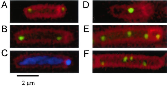
FIG. 4.
Visualization of chromosomal regions during sporulation and growth. (A to E) Cells were grown in defined minimal medium and induced to sporulate by the addition of mycophenolic acid. Samples were taken 4 h after the initiation of sporulation, and membranes were stained with FM4-64. The positions of origin regions were visualized with LacI-GFP bound to an array of lac operators inserted at 359°. Note that for cells with two or more foci, there was no detectable effect of spo0J on the frequency of positioning an origin region in the forespore (see text). Images illustrate some of the different types of sporangia observed. (A) Typical origin position in spo0J+ cells (strain PSL62 spo0J+ ΔspoIIIE::tet). (B to E) spo0J mutant cells [strain PSL73 Δ(soj-spo0J)::spc ΔspoIIIE::tet] with two origin foci located in the mother cell (B) and the chromosomal DNA visualized with DAPI (C), indicating that there is DNA in the forespore. (D) A spo0J mutant cell with a single focus of the origin region that is excluded from the forespore. (E) A spo0J mutant cell with four copies of the origin region, one of which is in the forespore. (F) Increased copies of the terminus region, visualized with LacI-CFP bound to an array of lac operators inserted at 181°, in a spo0J mutant (strain PSL110) during exponential growth.

