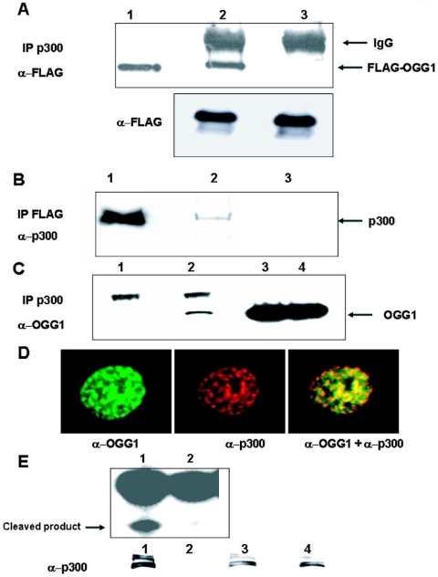
FIG. 1.
In vivo interaction of OGG1 and p300. A. Extracts of HCT116 cells cotransfected with expression plasmid for FLAG-tagged OGG1 and p300 were immunoprecipitated with p300 antibody (lane 2) or preimmune sera (lane 3) and then blotted with FLAG antibody. Lane 1, FLAG-tagged OGG1 in cell extract as marker. Lower panel, Western analysis with anti-FLAG antibody of cell extracts used in upper panel. B. Extracts of cells transfected with FLAG-tagged OGG1 were immunoprecipitated with FLAG antibody (lane 2) or preimmune sera (lane 3), and the immunoprecipitates were analyzed for p300 by Western blotting. Lane 1, cell extracts used as marker. C. Extracts of HCT116 cells were immunoprecipitated with p300 antibody (lane 2) or preimmune sera (lane 1) and then immunoblotted with OGG1 antibody; lanes 3 and 4, input controls of cell extracts. D. Colocalization of OGG1 and p300. MRC5 cells were immunostained with OGG1 (green) and p300 (red). E. Incision activity of p300 immunoprecipitates (lane 1) or preimmune sera (lane 2) with 32P-labeled 8-oxoG·C oligonucleotide. Lower panel, Western analysis of immunoprecipitates with p300 antibody and input controls of cell extracts (lanes 3 and 4).

