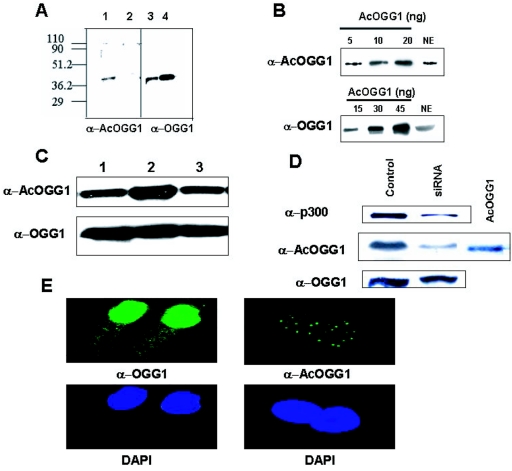
FIG. 4.
Identification and localization of AcOGG1 in vivo. A. Western analysis of in vitro-acetylated OGG1 (15 ng, lane 1) or unmodified OGG1 (45 ng, lane 2) with AcOGG1 antibody. Right panel, Western blot analysis of the same blot with OGG1 antibody (lanes 3 and 4). Numbers at left are molecular masses in kilodaltons. B. Western blot analysis of HeLa nuclear extract (75 μg) with AcOGG1 antibody (upper panel) or OGG1 antibody (lower panel) with known amounts of recombinant AcOGG1. NE, nuclear extract. C. Nuclear extracts (100 μg) of HCT116 cells transfected with pcDNA3 (lane 1) or WT p300 expression plasmid (lane 2) or p300 HAT mutant (lane 3) were immunoblotted with AcOGG1 antibody (upper panel) or OGG1 antibody (lower panel). D. HCT116 cells were transfected with 100 nM control siRNA (lane 1) or p300-specific duplex siRNA (lane 2); 48 h later, cell extracts were analyzed by Western blotting using p300 (upper panel)-, AcOGG1 (middle panel)-, or OGG1 (lower panel)-specific antibodies. E. Immunofluorescence studies of MRC5 cells with AcOGG1 antibody (right panel) or OGG1 antibody (left panel). Lower panels, cells were stained with DAPI.

