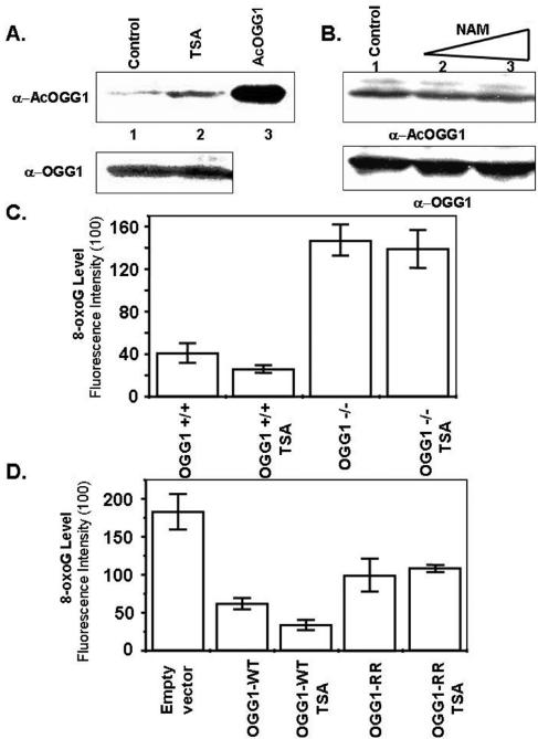
FIG. 7.
Effect of TSA on the level of AcOGG1 and 8-oxoG repair in OGG+/+ and OGG−/− MEF cells. A. HCT116 cells were either treated with TSA (100 ng/ml) for 12 h (lane 2) or mock treated (lane 1), and then cell extracts were immunoblotted with AcOGG1 (upper panel) or OGG1 (lower panel) antibody. Lane 3, AcOGG1 marker. B. HCT116 cells were treated with NAM (1 mM, lane 2; 5 mM, lane 3) for 12 h or mock treated (lane 1), and then cell extracts were immunoblotted with AcOGG1 (upper panel) or OGG1 (lower panel) antibodies. C. OGG1-null and WT MEFs were treated with TSA (12 h), and immunofluorescence of 8-oxoG in the genome was quantitated with 8-oxoG-specific antibody conjugated with fluorescein isothiocyanate. D. OGG1−/− MEFs were transfected with FLAG WT OGG1 or FLAG K338R/K341R mutant (OGG1 RR) or empty vector. Thirty-six hours after transfection, cells were mock treated or treated with TSA (12 h) and immunofluorescence of 8-oxoG was quantitated as before with fluorescein isothiocyanate. Other details are described in Materials and Methods.

