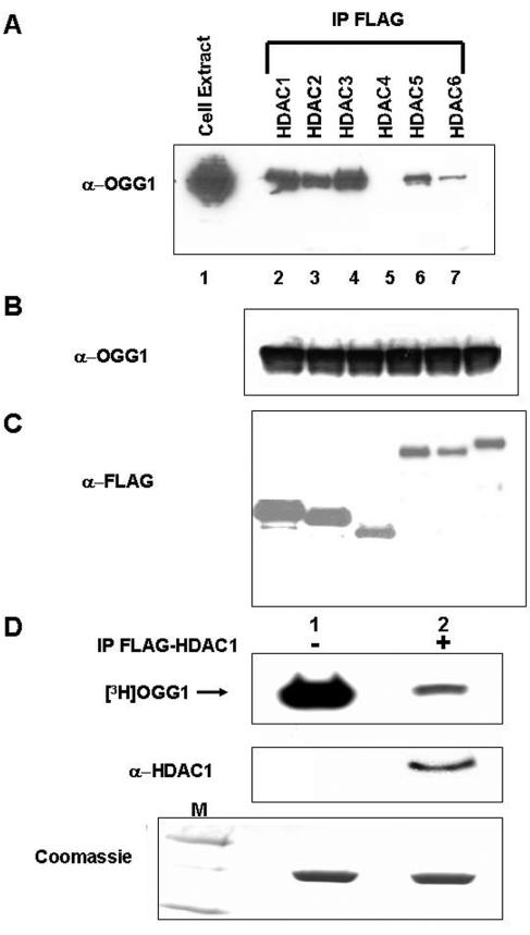
FIG. 9.
In vivo interaction of OGG1 and HDACs. A. HCT116 cells were transfected with expression plasmids for OGG1 and FLAG-tagged HDAC1 through HDAC6. FLAG immunoprecipitates of FLAG antibody were analyzed for OGG1. Lane 1, cell extract. B. Western analysis for OGG1. C. Western analysis for HDACs. D. In vitro-acetylated [3H]OGG1 (2 μg) was incubated with FLAG immunoprecipitates of FLAG-tagged HDAC1-transfected cells (lane 2) or empty vector-transfected cells (lane 1) at 30°C in HAT assay buffer for 45 min and analyzed by SDS-PAGE and fluorography. Middle panel, immunoblotting with HDAC1 antibody. Lower panel, Coomassie blue staining of input OGG1in a duplicate gel. Lane M, molecular weight markers.

