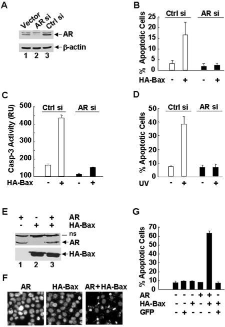
FIG. 2.
AR is required for UV- and Bax-induced apoptosis in prostate cancer cells. (A) 104-R1 cells were stably transfected with mammalian expression vectors encoding AR siRNA (AR si), the control scramble siRNA (Ctrl si), or empty vector. Expression levels of AR proteins were examined by immunoblotting with anti-AR antibody (AN21). (B and C) 104-R1 (AR si and Ctrl si) cells were infected with Ad/Bax plus Ad/Cre (HA-Bax +) or Ad/Bax plus Ad/Luc (HA-Bax −) for 24 h. The apoptotic cell death was then detected and quantitated by Hoechst (H33258) staining (B) or by caspase activity assays with the fluorogenic substrate DEVD-AFC (C). (D) Cells were treated with UV (10 mJ/cm2) for 18 h. The apoptotic cell death was detected as described in panel B. (E, F, and G) AR-negative PC3 cells were transiently transfected with an expression vector encoding AR or green fluorescent protein (GFP). After 20 h, cells were infected with Ad/Bax plus Ad/Cre for another 16 h. (E) Expression of AR and HA-Bax proteins was analyzed by immunoblotting with antibodies against AR or HA, respectively. (F) The apoptotic cells were examined by nuclear staining with Hoechst (H33258) and visualized under UV fluorescence microscope. (G) Apoptotic cells were also quantitated by counting immunostaining AR-positive cells, which also have apoptotic nuclei as shown by staining with Hoechst stain (H33258). GFP-positive cells were quantitated in the same manner.

