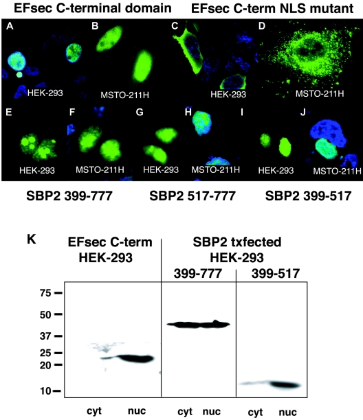
FIG. 3.
EFsec and SBP2 contain functional NES and NLS signals. (A and B) Localization of transiently expressed FLAG-tagged EFsec C-terminal domain in HEK-293 (A) and MSTO-211H (B) visualized by confocal microscopy using 1:500 anti-FLAG/1:500 anti-mouse antibody-FITC. (C and D) EFsec C-terminal domain NLS mutant (K536E, K537E) in HEK-293 (C) and MSTO-211H (D) cells stained as described for panel A. (E to J) Localization of mutant SBP2 constructs in HEK-293 (E, G, and I) and MSTO-211H (F, H, and J) cells visualized by immunostaining with 1:500 anti-V5/1:500 anti-mouse antibody-Alexa Fluor 488. Panels E and F indicate SBP2 399-777-V5 (minimal functional domain). Panels G and H indicate SBP2 517-777-V5 (transactivation domain). Panels I and J indicate SBP2 399-517-V5 (SECIS-RNA binding domain). In some panels, blue staining with 1:1,500 DAPI indicates nucleus of the cell. Cyan areas indicate the merging of blue and green fluorescence. (K) Subcellular fractionation and localization of EFsec C-terminal domain, SBP2 399-777, and SBP2 399-517 was carried out as described in Materials and Methods. Numbers on the left side of the panel indicate molecular masses in kilodaltons. Txfected indicates transfected. cyt, cytoplasmic extract; nuc, nuclear lysate.

