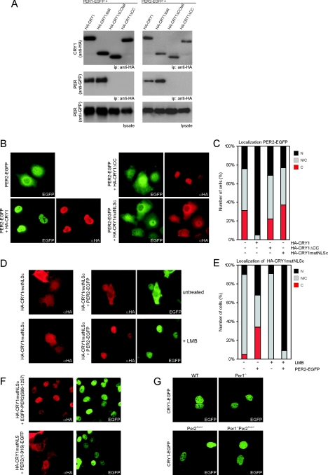
FIG.2.
Subcellular localization equilibrium of the mCRY1-mPER2 complex. (A) Western blot of an immunoprecipitation (ip) of COS7 cells transfected with HA-CRY1 (wild type or mutant) and either PER1-EGFP (left) or PER2-EGFP (right). Proteins were precipitated from the lysate with anti-HA antibodies and then analyzed on Western blots using anti-HA (top panels) and anti-GFP (middle panels) antibodies. The bottom panels show Western blots of the total lysate using anti-GFP antibody. (B) Immunofluorescence pictures of COS7 cells transfected with PER2-EGFP alone or with PER2-EGFP and HA-CRY1, HA-CRY1ΔCC, or HA-CRY1mutNLSc. αHA, anti-HA. (C) Quantification of the cellular localization of PER2-EGFP shown in panel B. (D) Immunofluorescence pictures of COS7 cells transfected with HA-CRY1mutNLSc alone (top panels) or with HA-CRY1mutNLSc and PER2-EGFP (bottom panels). Treatment of cells with LMB is indicated (+ LMB). (E) Quantification of the cellular localization of HA-CRY1mutNLSc, as shown in panel D. (F) Immunofluorescence pictures of COS7 cells transfected with HA-CRY1mutNLSc and EGFP-PER2(596-1257) (top) or PER2(1-916)-EGFP (bottom). (G) Fluorescence microscopy pictures of wild-type (WT), Per1−/−, Per2Brdm1/Brdm1, and Per1−/− Per2Brdm1/Brdm1 MDFs transiently expressing mCRY1-EGFP.

