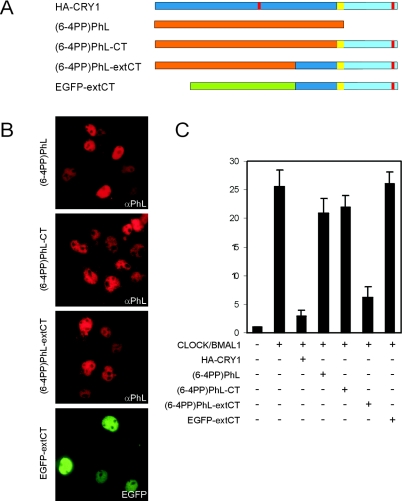
FIG. 4.
Cross talk between the photolyase-like domain of mCRY1 and its C terminus. (A) Schematic representation of HA-CRY1, (6-4PP)PhL, and the fusion proteins (6-4PP)PhL-CT, (6-4PP)PhL-extCT, and EGFP-extCT. (B) Immunofluorescence pictures of transfected COS7 cells showing the subcellular localization of the aforementioned proteins. αPhL, anti-PhL. (C) Graphic representation of the CLOCK/BMAL1-inhibitory capacity of the proteins in the Dbp E-box promoter-luciferase reporter assay (using 100 ng of plasmid). Error bars represent the standard deviations. The difference between HA-CRY1 and (6-4PP)Phl-extCT is not statistically significant (t test P value is 0.07).

