FIG. 6.
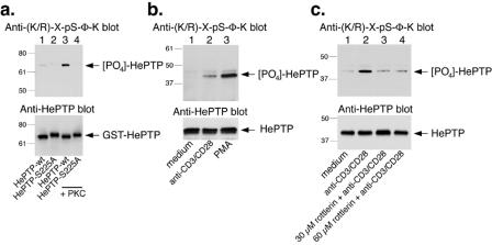
PKC θ phosphorylates HePTP at S225 in primary T cells. (a) Upper panel, anti-(K/R)-X-pS-Φ-K immunoblot of GST-HePTP (lanes 1 and 3) and GST-HePTP-S225A (lanes 2 and 4) incubated with kinase buffer alone (lanes 1 and 2) or with PKC (lanes 3 and 4). Lower panel, anti-HePTP blot of the same filter. (b) Upper panel, anti-(K/R)-X-pS-Φ-K immunoblot of anti-HePTP immunoprecipitates from primary human T cells left untreated (lane 1) or stimulated with anti-CD3 and anti-CD28 Dynabeads (lane 2) or 50 nM phorbol ester (lane 3) for 10 min before lysis. Lower panel, anti-HePTP immunoblot of the same immunoprecipitates. (c) Upper panel, anti-(K/R)-X-pS-Φ-K immunoblot of anti-HePTP immunoprecipitates from primary human T cells left untreated (lane 1), stimulated with anti-CD3 (lane 2), or treated with 30 μM rottlerin plus anti-CD3/CD28 beads (lane 3) or with 60 μM rottlerin plus anti-CD3/CD28 beads (lane 4). Lower panel, anti-HePTP immunoblot of the same immunoprecipitates. Mrs (in thousands) are shown to the left of the gels.

