FIG.4.
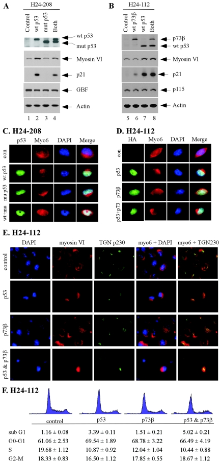
Intracellular localization of myosin VI is altered by wild-type p53, but not mutant p53 or wild-type p73β. (A) Myosin VI is induced by wild-type (wt) p53, but not mutant (mut) p53(R273H). H24-208 cells were uninduced (control), induced to express wild-type p53, induced to express mutant p53(R273H), or induced to express both for 48 h. The expression level of wild-type p53, mutant p53, myosin VI, p21, and GBF was quantified by Western blot analysis. Actin was analyzed as a loading control. (B) Myosin VI is induced by wild-type p53, but not wild-type p73β. H24-112 cells were uninduced (control), induced to express wild-type p73β, induced to express wild-type p53, or induced to express both for 48 h. The expression level of p73β, p53, myosin VI, p21, and p115 was quantified by Western blot analysis. (C) The intracellular localization of myosin VI is altered by wild-type p53, which is inhibited by mutant p53(R273H). Immunofluorescence staining was performed as described in Materials and Methods. p53 was stained green, whereas myosin VI was stained red. Nuclei were stained blue. con, control. (D) The intracellular localization of myosin VI is altered by wild-type p53, but not wild-type p73β. (E) Colocalization of myosin VI with trans-Golgi marker TGN p230. (F) Cell cycle arrest is not sufficient to alter cellular localization of myosin VI. H1299 cells were uninduced or induced to express p53, p73β, or both. The cell cycle profile was determined by DNA histogram analysis 36 h following induction of p53, p73, or both.

