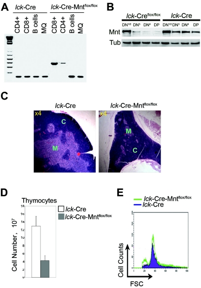
FIG. 1.
Deletion of Mnt in T cells causes decreased thymic cellularity. (A) PCR genotyping performed on DNA obtained from FACS-sorted thymic CD4+ and CD8+ T cells, splenic B220+ B cells, and F4/80+ macrophages from mice of the indicated genotypes at 8 weeks of age. The 160-bp PCR product is diagnostic for the wild-type Mnt allele, and the 350-bp PCR product is diagnostic for the mutant allele. (B) Western blot analysis of Mnt expression in DN subsets obtained from lck-Cre Mntflox/flox and lck-Cre mice. DN T cells were enriched from total thymocytes isolated from multiple 5-week-old littermates by first removing populations staining with antibodies against CD3, CD4, CD8, GR1, Mac1, and B220. DN T cells were then sorted by flow cytometry for CD25 and CD44 to isolate the different DN subsets. The DN1 and DN2 populations were pooled because of their relatively low numbers. Tub, tubulin. (C) Hematoxylin-and-eosin-stained thymus sections. The inner medulla (M) region of the lck-Cre Mntflox/flox thymus was markedly reduced relative to the cortex (C). Magnification (n-fold) is indicated. (D) Comparison of total thymocyte numbers from lck-Cre-Mntflox/flox mice (n = 5) and age-matched (5- to 9-week-old) control mice (n = 6). Values shown are means ± standard deviations (P = 0.005). (E) Cell size comparison of total thymocytes determined by FSC.

