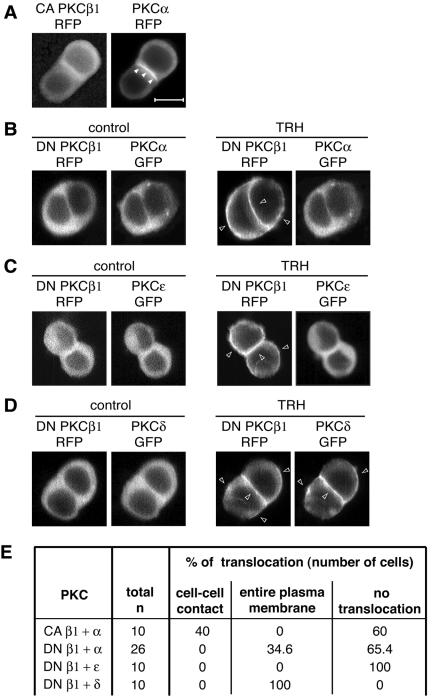
FIG. 5.
PKCα and -ɛ translocation depends on PKCβ1 activity. CA PKCβ1-RFP was first cotransfected with PKCα-GFP in GH3B6 cells, and observations were performed 48 h later. (A) PKCα-GFP still accumulated at cell-cell contacts in the presence of CA PKCβ1, which bypasses receptor activation and therefore does not induce any increase in intracellular Ca2+ and DAG concentrations. Observations were performed with a conventional microscope. A video in the supplemental material shows that CA PKCβ1-GFP oscillates spontaneously, without TRH, between the cytoplasm and plasma membrane. In the presence of DN PKCβ1-RFP (B) and in the presence of 100 nM TRH, PKCα-GFP remained in the cytoplasm of most cells or accumulated at the entire plasma membrane in 9/26 cells, as did PKCɛ (C). PKCδ translocation was not affected by the cotransfection of DN PKCβ1-RFP (D). All the data presented in panels A to D were obtained 48 h after the cotransfection by real-time recordings. (E) Summary of the total number of cells analyzed in each situation and the subcellular localizations of the different GFP fusion proteins. As previously shown by others for DN PKCγ and -δ (49), we found that in the presence of 100 nM TRH, DN PKCβ1 accumulated at the same site as the native enzyme, namely, at the entire plasma membrane (B, C, and D), while the CA PKCβ1 keeps oscillating between the cytoplasm and plasma membrane (see the video in the supplemental material). In each experiment, it was checked (n = 5 cells for each PKC) that PKCα, PKCɛ, and PKCδ-GFP transfected alone were localized exclusively at cell-cell contacts.

