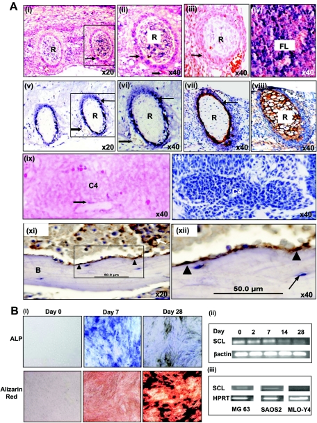
FIG. 1.
(A) SCL is expressed in bone. (i) Section through ribs of an E14.5 embryo hybridized with an SCL antisense probe. SCL RNA is expressed in the perichondrium (arrow) of ribs (R). (ii) Magnified view of boxed area in panel i showing RNA expression in the perichondrium (arrow) and endothelium (block arrow) of blood vessels. (iii) Tissue section corresponding to panel i, probed with a sense SCL probe as a negative control. (iv) Section through fetal liver (FL) showing SCL expression in blood progenitors. (v and vi) Immunohistochemical studies of SCL expression in E14.5 mouse embryos with SCL-positive cells stained blue-black. (v) Ribs in cross section showing expression of SCL in cells in the perichondrium (arrow) and endothelial cells (block arrow). (vi) Magnified view of boxed area in panel v. (vii and viii) Immunohistochemical staining of osteoblasts and chondrocytes in rib sections of E14.5 mouse embryos. (vii) Osteoblasts (detected using a collagen 1 antibody and stained brown) are seen predominantly in the perichondrium (arrow). (viii) Chondrocytes (detected using a collagen 2 antibody and stained brown) fill the cartilaginous template. (ix) Section through the centrum of the C4 vertebra in an E11.5 mouse embryo hybridized with an SCL antisense probe. SCL RNA is expressed in endothelium (block arrow) but not in the vertebral template. (x) An E11.5 section corresponding to panel ix, stained for osteoblasts using a collagen 1 antibody. Collagen 1-expressing osteoblasts are also not present at this stage. (xi and xii) Immunohistochemical studies of SCL expression in adult human bone marrow with SCL-positive cells stained brown. (xii) SCL was expressed by osteoblasts (arrowhead) lining the endosteal surface of bone (B) and a subset of hematopoietic cells (arrow). (xiii) Magnified view of the boxed area in panel xi showing SCL expression in osteoblasts (arrowhead) but not in osteocytes (arrow). (B) SCL expression falls with bone differentiation. (i) Osteogenic differentiation of the preosteoblast cell line MC3T3-E1. Cells cultured in bone differentiation medium were stained at days 0, 7, and 28 for alkaline phosphatase activity (ALP, upper panel) and alizarin red staining (lower panel). Alkaline phosphatase activity (intense blue) was prominent at day 7 and decreased with the acquisition of a mature osteocytic phenotype at day 28. There was a corresponding increase in the degree of matrix mineralization as assessed by alizarin red staining (intense red) from days 0 to 28. (ii) SCL expression in MC3T3 cells was assessed by RT-PCR at various time points and decreased with bone differentiation. (iii) SCL expression in the osteoblastic cell lines MG63 and SAOS2 and the osteocytic cell line MLO-Y4 was assessed by RT-PCR. SCL was expressed in the osteoblastic cell lines but was absent in MLO-Y4 cells, which have the phenotype of terminally differentiated osteocytes.

