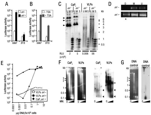
FIG. 1.
High-level, copy number-independent VLP-delivered transgene expression activated by viral factors or TSA. (A and B) Luciferase activity (RLU/mg total protein) from (A) cos7 or 3T3 cells treated with VLPs carrying pDNA without (−) or with (+) ori and from (B) cos7 or 3T3 cells treated with VLPs carrying pDNA without ori, cultured without or with TSA. Note: mock VLP-treated cells cultured with TSA resulted in less than 100 RLU/mg total protein (data not shown); y axis is a log scale. Error bars indicate the standard errors. (C) Southern blot analysis of low-molecular-weight DNA Hirt extracts (26) from cos7 cells transfected using CaPi or treated with VLPs carrying pDNA without (−) or with (+) ori. Restriction endonuclease digests of DNA extracts (U, mock-digested DNA; M, MboI [inhibited by dam methylation, digests only DNA replicated in mammalian cells]; or S, Sau3A [digests all DNA]) demonstrated pDNA replication products present in CaPi-transfected but not VLP-treated cells (indicated by asterisks). The positions of migration of form I, II, and III DNAs are indicated by arrowheads. Considerable degradation is observed for the VLP-delivered pDNA, presumably due to the majority of the plasmid being unprotected from nuclease attack in the cell (52). Luciferase activity (RLU/mg total protein) from an aliquot of each transfected cell culture taken prior to Hirt extraction is given below the appropriate tracks. Positions of migration of molecular weight standard markers are shown to the right of the gel. (D) PCR analyses of low-molecular-weight DNA extracts from cos7 cells treated with VLPs as described for panel C. Plasmid-specific PCR amplification was performed after digestion of the extracts with the restriction endonucleases DpnI (D, inhibited by mammalian methylation; PCR amplification detects mammalian replicated pDNA only; indicated by asterisks) or MboI (M, inhibited by dam methylation; PCR amplification detects bacterial input pDNA only) or mock-digested DNA (U). (E) Luciferase activity from cos7 cells treated with increasing amounts of VLPs or CaPi (equivalent to 0.0005 to 5 μg pDNA/5 × 104 cells) carrying pDNA without (−) or with (+) ori. Note: both axes are a log scale. (F) Micrococcal nuclease assays of HeLa cell nuclei transfected with CaPi or treated with VLPs. Following electrophoresis of micrococcal nuclease (MN)-treated samples, the agarose gels were stained with ethidium bromide (left panels) and then subjected to Southern blot analysis (right panels). Consistent with previous observations (29), atypical nucleosome ladders for CaPi-transfected pDNA were detected, represented by protected pDNA fragments of approximately 0.1 and 0.6 kb (arrowheads). Similar ladders were detected for VLP-delivered pDNA. A further band of VLP-protected pDNA fragments (52) of 1.2 to 1.5 kb was also detected (asterisks). (G) Untransfected HeLa cell nuclei with pDNA added to isolated nuclei prior to micrococcal nuclease digestion and then analyzed as described above for panel F. Left panel, ethidium bromide stain; right panel, Southern blot analysis. Positions of migration of molecular weight standard markers are shown to the left of the gels.

