
Figure 4.
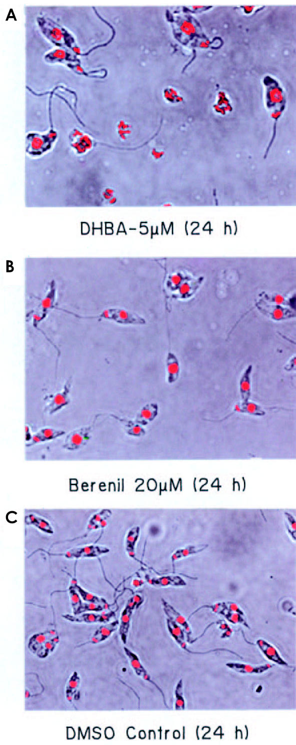
Confocal microscopy of L. donovani AG 83 promastigotes. Parasites incubated with 5 μM DHBA (A), 20 μM berenil (B) and 0.5% DMSO (C) were stained with ethidium (0.1 μg/mL) in 1× phosphate-buffered saline containing 10% glycerol. The cells were viewed under a Leica DM IRB inverted microscope. Phase contrast and fluorescence images for the same fields were merged and are represented here. Magnification 1000×.