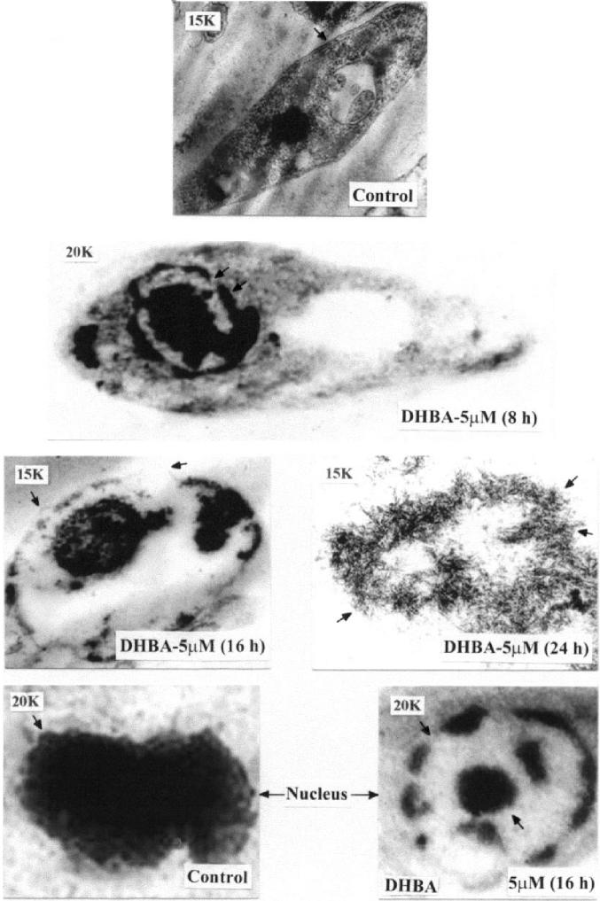
Figure 5.
Transmission electron microscopy of DHBA-treated promastigotes. L. donovani AG83 cells were cultured in media containing 0.5% DMSO or 5 μM DHBA for different time periods. Spurr blocks were prepared as described in Materials and Methods. Control cells treated with 0.5% DMSO; cells treated with 5 μM DHBA for 8 hr (arrow shows cup-shaped masses); cells treated for 16 h (arrows show surface blebbing); cells treated for 24 h (arrows show apoptotic bodies); nucleus of control cell (arrow shows intactness of nucleus); nucleus of cells treated with 5 μM DHBA for 16 h (arrows indicate chromatin margination). Magnification of 15000 (15K) and 20000 (20K) are indicated in each figure.

