Figure 1.
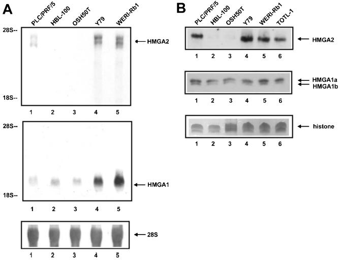
High-level expression of HMGA2 and HMGA1 mRNA and protein in cultured retinoblastoma cells. A: Northern blot of total RNA isolated from the Y79 (lane 4) and WERI-Rb1 (lane 5) retinoblastoma cells, PLC/PRF/5 hepatoma cells (lane 1), HBL-100 mammary epithelial cells (lane 2), and OSH50T osteosarcoma cells (lane 3), probed, respectively, with the radiolabeled HMGA2 and HMGA1 coding region DNA (upper and middle panels). 28S and 18S rRNA provide size markers of 4.8 and 1.8 kb. Uniform RNA loading in individual sample was shown by hybridizing the same Northern blot with the 28S rRNA probe (lower panel). B: Western blot of perchloric acid extracts from cells of PLC/PRF/5, HBL-100, OSH50T, Y79, and WERI-Rb1 (lanes 1 to 5), and an additional retinoblastoma cell line TOTL-1 (lane 6); cells were prepared and equal protein loading between lanes confirmed by the histone H1 band stained by Coomassie (lower panel). Anti-HMGA2 and A1 polyclonal antibodies were used to detect HMGA2 (upper panel) and HMGA1a/b (middle panel) proteins.

