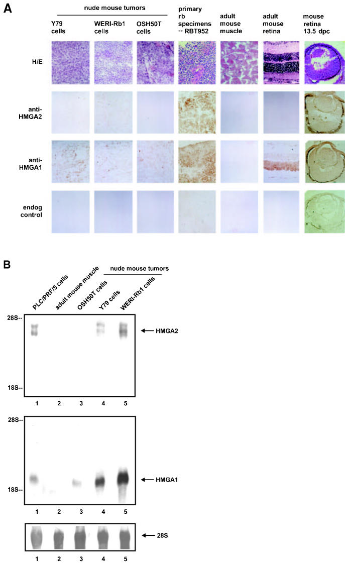
Figure 2.
HMGA2 expression is in established lines of retinoblastoma cells propagated in nude mice, surgically removed retinoblastoma samples, and mouse embryonic retina, but absent in adult mouse tissues. A: Immunohistochemistry of cryosections of nude mouse tumors derived from the retinoblastoma cells Y79 and WERI-Rb1, and that from osteosarcoma cells OSH50T were hematoxylin & eosin stained, immunoperoxidase stained with the polyclonal antibodies specifically recognizing only HMGA2 protein (anti-HMGA2 panel), immunostained with the polyclonal antibodies which recognize only HMGA1 protein (anti-HMGA1 panel), and endogenous peroxidase controlled (endog control panel). HMGA proteins were detected in the primary retinoblastoma sample RBT952 (see Figure 3) as well. Retina of 129/sv mouse embryo 13.5-d postcoitum showed positive HMGA2 and HMGA1 immunostaining, whereas 6-wk-old Balb/c retina demonstrated undetectable HMGA2 protein but abundant HMGA1 protein. Thigh muscle from adult nude mouse did not stain with HMGA2 or HMGA1. Photomicrographs are magnified 400×. B: Northern blot of total RNA prepared from PLC/PRF/5 hepatoma cells (lane 1), adult mouse thigh muscle (lane 2), and nude mouse tumors derived respectively from OSH50T (lane 3), Y79 (lane 4), and WERI-Rb1 (lane 5), probed separately with radiolabeled HMGA2 (upper panel) and HMGA1 (middle panel) coding region DNA, and 28S rRNA (lower panel).

