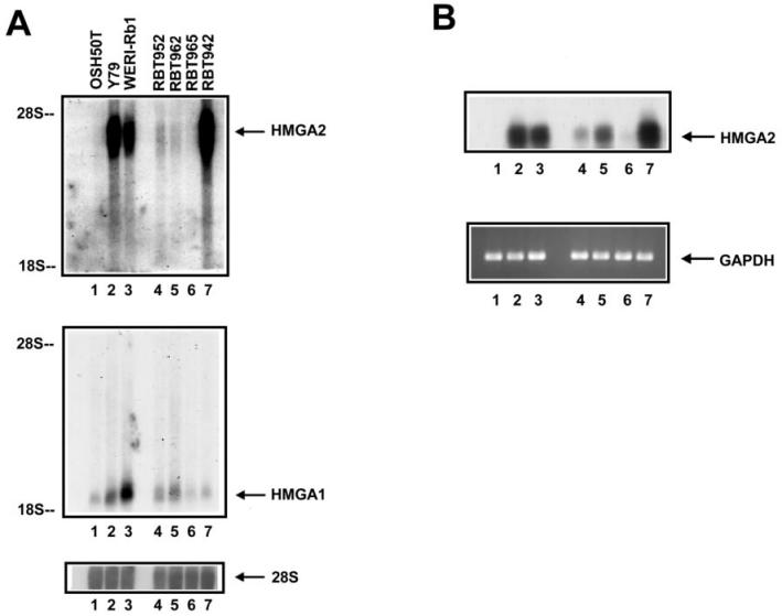
Figure 3.
High prevalence of HMGA2 gene expression in surgically removed retinoblastoma samples. Intact RNA was prepared from the cells: OSH50T (lane 1), Y79 (lane 2) and WERI-Rb1 (lane 3), and the surgically removed retinoblastoma specimens: RBT952, RBT962, RBT965, and RBT942 (lanes 4 to 7). A: Northern blot of the HMGA2 (upper panel) and HMGA1 (middle panel) mRNA, in which equal RNA loading was controlled by 28S probing (lower panel). Autoradiograph of the upper panel was overexposed to reveal weak signals. B: RT-PCR analysis of the same RNA samples, in which reverse transcription was performed separately using a gene-specific primer for HMGA2 and random primers. cDNA synthesized were used in PCR amplification with pairs of HMGA2 and GAPDH-specific primers. To ensure amplification is specific, PCR products from HMGA2 gene amplification were size fractionated onto an agarose gel, blotted onto a nylon membrane and hybridized with HMGA2 probe (upper panel). PCR products from GAPDH amplification, electrophoresed through an agarose gel and stained with ethidium bromide (lower panel), showed the equal quantities of cDNA used.

