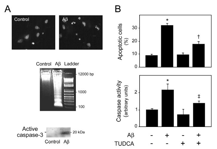
Figure 1.
Aβ-induced apoptosis in rat cortical neurons is inhibited by TUDCA. Cells cultured for 3 d were incubated either with vehicle (control), 25 μM Aβ fragment 25–35, 100 μM TUDCA, or a combination of Aβ plus TUDCA for 24 h. In co-incubation experiments, cells were pretreated with TUDCA for 12 h and the bile acid was left in the culture medium with Aβ. A: Morphological and biochemical characteristics of apoptosis in rat cortical neurons incubated with Aβ peptide. Fluorescence microscopy of Hoechst staining (top) shows condensed or fragmented nuclei indicative of apoptosis. DNA laddering (middle) and caspase-3 processing (bottom) are also characteristic of apoptotic cell death. B: TUDCA prevents Aβ-induced apoptosis in rat cortical neurons. Histograms show mean ±SEM values of nuclear fragmentation (top) and caspase-3–like activity (bottom) for at least 5 independent experiments. *P < 0.01 from control; †P < 0.01 and ‡P < 0.05 from Aβ peptide.

